Friday, November 8, 2019

A Way Out


A majority of our life is defined by our choices. For the most part, we have the power to decide everything from what we want to eat today to what we want to spend the rest of our lives doing. But what about those life-altering decisions that have already been made for us, sometimes even before we are born? Is it just the luck of the draw? Should we just accept the cards we were dealt with or is there something we can do to  change the course of our lives?

        For years, many have succumbed to incurable diseases because the therapies used to treat the disease were anything but sufficient. People worldwide suffer from inherited disorders that can cost them later in life and even impact the future of generations to come. One such disease is hypertrophic cardiomyopathy (HCM). HCM is a genetic disorder that displays an autosomal dominant inheritance pattern1. According to the National Human Genome Research Institute, autosomal dominant explains how certain traits are inherited. Autosomal signifies that the trait acquired is from a non-sex chromosome; thus, for humans, autosomal diseases are derived from chromosomes 1 through 22. Furthermore, dominant explains how many copies of the mutated gene are required for the disease phenotype. If it is dominant disorder, then only one copy of the mutated gene will display the disease phenotype. Other examples of autosomal dominant diseases are Huntington disease and Marfan Syndrome2.

        Therefore, in cases of familial hypertrophic cardiomyopathy, it only takes one mutated
gene to dictate what the rest of your life will look like. HCM is depicted by the thickening of the left ventricle of the heart, lowered chamber capacity, and an enlarged heart1. The muscle walls of heart thicken which restricts blood flow causing the ventricle to work harder to pump blood to the rest of the body4. The prevalence of HCM is estimated to occur in around 1 out of 625 people to 1 in 344 people1. The European Society of Cardiology suggests that a heart wall thickness greater than or equal to 15 mm is indicative of HCM3.  HCM is a hereditary disorder which affects approximately 0.2% of the population worldwide1. Common symptoms of HCM include dizziness, fainting, fatigue, chest pain, shortness of breath, and arrhythmias which can lead to death7.
        While a number of different mutational events can occur, the most prevalent, accounting for up to 40% of HCM cases involve mutations in a gene called MYBPC3.  This gene is responsible for creating the cardiac myosin binding protein C, a cardiac protein, and is typically inherited from at least one parent8. Treatment of patients who have MYBPC3 mutations involve regular monitoring and medications and to open heart surgery or an implantable cardioverter-defibrillator (ICD)5. While these treatments options will only alleviate symptoms and are temporary solutions, there is no cure presently. The survival rate of people with HCM is 98% after 1 year, 94.3% after 3, and 82.2% after 56. While HCM is not necessarily a death sentence, a person’s mortality is always looming over their shoulder. People should not have to live in fear, wondering which day will be their last. Perhaps with genome editing, they won’t have to anymore.
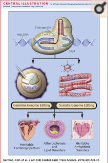
        In a 2017 study conducted by Ma et al, the effects of genome editing to correct germline mutations that cause HCM were explored. Here, healthy gamete donors who had homozygous and heterozygous heritable MYBPC3 mutations were used. They used CRISPR-Cas9 to specifically target the heterozygous MYBPC3 mutation in embryos prior to implantation8. CRISPR-Cas9 is a genome editing technology that allows for an organism’s DNA to be altered. The technology involves creation of a small piece of RNA which contains a guide sequence that attaches to a specific portion of the DNA. That RNA piece will also bind to an enzyme called Cas9. Essentially, the RNA recognizes the portion of DNA which needs to be removed from the genome and Cas9 will cut that DNA at the specific location. After the DNA is cut, a customized DNA sequence will replace the sequence that was removed from the genome9. Therefore, this technology is especially promising for individuals with mutations that cause incurable diseases. 

        Ma et al. explains that homologous direct repair (HDR) is necessary for gene correction. They found that the double strand breaks in the human gametes and zygotes were fixed by using an internal HDR mechanism by using a wild-type allele as a template. However, in induced pluripotent stem cells, the rate of HDR was much lower indicating that DNA damage response system behaved differently in gametes and embryos. One problem, though, is that , there are still off target effects like non-homologous end joining induced indels that need to be controlled for and understood8. We are not close to where we need to be, but this is one step to finding the answer. Undoubtedly, there are many challenges to using gene therapy. The first of which is limited sample size and the difficulty in setting clear boundaries10. Furthermore, editing the germline can cause intended consequences to future generations. Although there is heavy criticism and controversy surrounding this study, I am hopeful for the future. Right now, CRISPR-Cas9 has only been tested on animal models and embryos, but in the future, I expect to see this technology being used in humans who discover their diseases later in life.  After more testing, the goal is to help those who are victims to genetic mutations and allow them to live disease free. If this expands to humans and is successful, individuals will no longer have to succumb to the whimsy of their genes.

By Rachel Crasta, Master of Medical Sciences Student, University of Kentucky

References:

1.      Marian, Ali J., and Eugene Braunwald. “Hypertrophic Cardiomyopathy.” Circulation Research, vol. 121, no. 7, 2017, pp. 749–770., doi:10.1161/circresaha.117.311059.
2.      “Autosomal Dominant.” Genome.gov, NIH, www.genome.gov/genetics-glossary/Autosomal-Dominant.
3.      Authors/Task Force Members, Elliott PM, Anastasakis A, Borger MA, Borggrefe M, Cecchi F, Charron P, Hagege AA, Lafont A, Limongelli G, Mahrholdt H, McKenna WJ, Mogensen J, Nihoyannopoulos P, Nistri S, Pieper PG, Pieske B, Rapezzi C, Rutten FH, Tillmanns C, Watkins H. 2014 ESC guidelines on diagnosis and management of hypertrophic cardiomyopathy: the task force for the diagnosis and management of hypertrophic cardiomyopathy of the european society of cardiology (ESC). Eur Heart J. 2014;35:2733–2779. doi: 10.1093/eurheartj/ehu284.
4.      “Hypertrophic Cardiomyopathy.” Www.heart.org, www.heart.org/en/health-topics/cardiomyopathy/what-is-cardiomyopathy-in-adults/hypertrophic-cardiomyopathy.
5.      Mcnamara, James W., et al. “MYBPC3 Mutations Are Associated with a Reduced Super-Relaxed State in Patients with Hypertrophic Cardiomyopathy.” Plos One, vol. 12, no. 6, 2017, doi:10.1371/journal.pone.0180064.
            6.    Liu, Qun, et al. “Survival and Prognostic Factors in Hypertrophic Cardiomyopathy: a Meta-  
                  Analysis.” Nature News, Nature Publishing Group, 20 Sept. 2017,                          
                  https://www.nature.com/articles/s41598-017-12289-4.
7.      “Hypertrophic Cardiomyopathy.” HIE Multimedia - Hypertrophic Cardiomyopathy, slu.adam.com/content.aspx?productId=117&isArticleLink=false&pid=1&gid=000192.
8.      Ma, Hong, et al. “Correction of a Pathogenic Gene Mutation in Human Embryos.” Nature, vol. 548, no. 7668, 2017, pp. 413–419.,doi:10.1038/nature23305.
9.      “What Are Genome Editing and CRISPR-Cas9? - Genetics Home Reference - NIH.” U.S. National Library of Medicine, National Institutes of Health, ghr.nlm.nih.gov/primer/genomicresearch/genomeediting.
10.  Papasavva, Panayiota, et al. “Rare Opportunities: CRISPR/Cas-Based Therapy Development for Rare Genetic Diseases.” Molecular Diagnosis & Therapy, vol. 23, no. 2, 2019, pp. 201–222., doi:10.1007/s40291-019-00392-3.


Monday, November 4, 2019

In Mice and Men (and Women): Prostate Drug, Terazosin to Slow Progression of Parkinson’s Disease


        In 1817, English physician James Parkinson published “An Essay on the Shaking Palsy,” which was the earliest known comprehensive explanation of Parkinson’s Disease (PD). Over 200 years later the underlying basis causing PD remains a mystery. However, research suggests that PD is a consequence of the complex, multifaceted relationship between several environmental and genetic components.1 Leucine-rich repeat kinase 2 (LRRK2) is a kinase enzyme encoded by the LRRK2 gene. Mutations or variations of the LRRK2 gene have been deemed as the principle factor manipulating the genome in most cases of familial PD, and some cases of sporadic PD.1 Hallmark signs and symptoms of PD (Figure 1.) are typically motor-related, for instance, the most common PD motor symptoms include bradykinesia, rigidity, resting tremor, stooped posture/unstable balance.1,2
Figure 1. Hallmark Symptoms and Signs of PD.2

        The basal ganglia system plays a major role in the initiation of movements (motor planning) and muscle tone. Two of the major nuclei found in the basal ganglia are the substantia nigra and the striatum. The striatum is further divided into three subsections, the caudate nucleus, putamen, and nucleus accumbens. Together, the caudate nucleus and putamen form the dorsal striatum. The caudate nucleus receives afferent signals from the prefrontal cortex, and it is responsible for cognitive function and regulating eye movements. On the other hand, the putamen receives afferent inputs from the motor cortex and the substantia nigra and it controls motor function, specifically voluntary movement. Another key nucleus of the basal ganglia system is the substantia nigra. Neurons of the substantia nigra use dopamine as a neurotransmitter, giving this area its unique distinguishing appearance.
        The substantia nigra appears unusually dark even in the absence of stain; this distinctive characteristic is a result of neuromelanin. Neuromelanin is a dark pigment formed during dopamine metabolism and is continuously produced by the densely packed dopamine neurons of the substantia nigra. These dopaminergic neurons are projected from the substantia nigra to the putamen region of the striatum, these afferent fibers are known as nigrostriatal fibers.
        Parkinson’s Disease progressively affects the nervous system, slowly degenerating and destroying dopaminergic neurons in the substantia nigra. As PD advances fewer neurons can be found in the substantia nigra which means that less dopamine is being released into the striatum. The neurodegenerative effects and dysfunction of the nigrostriatal striatal system observed in PD patients are direct consequences of the striatal dopamine-deficiency. 4,5 The absence of striatal dopamine results in the inability to conduct controlled, fine muscle movements.     
         Since there is no definitive diagnostic or imaging test to diagnose PD, diagnosis is made clinically. A clinical diagnosis of PD is only possible once the patient starts to exhibit symptoms of the disease. Once symptoms begin to manifest this indicates that the disease has progressed to a point where over 60% of nigral nerve cells have disappeared. 1 Every case of PD progresses differently, the disease may differ in severity, rate of progression, and an individual may experience different PD symptoms, presentation of neuropathology, age of onset, etc. An example of the average or general progression of PD and the clinical symptoms associated with each stage is shown in Figure 2.
Figure 2. Clinical symptoms associated with PD and general disease progression.3
In many cases, after the patient succumbs to the disease their brain can be examined for conclusive evidence of PD. For example, by immunohistochemically staining the substantia nigra of a PD patient, researchers can establish the extent of neuronal degeneration by comparing the section to the immunohistochemically stained substantia nigra from a healthy patient. 4,5
Unlike the healthy substantia nigra which consists of distinct deeply stained regions, a PD patient’s substantia nigra will show much lighter regions, ultimately allowing researchers to define the disease due to the observed depigmentation (Figure 3).3 
Figure 3. Main neuropathologies observed in immunohistochemical stains of PD patients.3 
In addition to the above-mentioned depigmentation neuropathological hallmark of Parkinson’s Disease is the presence of Lewy Bodies. Lewy Bodies are intracytoplasmic inclusions containing aggregations of the α‑synuclein protein.1,3 Lewy Bodies are known to cause mitochondrial dysfunction by disrupting the electron transport chain which leads to the degradation of dopaminergic neurons in adulthood. Damage to the mitochondria causes diminished ATP levels in PD patients because of their impaired cerebral glucose metabolism and mitochondrial biogenesis. Cellular dysfunctions, including diminished ATP levels and impaired bioenergetics, have been regularly observed in PD patients and may determine the severity and course of the disease.6 As PD progresses, the abnormal deposition and aggregation of the α‑synuclein protein within cytoplasm gradually increase and begin to involve more brain regions.7 Shown in Figure 4 are examples of immunohistochemical staining of three unique Lewy bodies all differing in their morphological characteristics.
Figure 4. Immunohistochemical staining of α -synuclein shows characteristic Lewy bodies.3

    The gold standard for Parkinson’s Disease drug therapy is L-DOPA, also called levodopa. Systemic administration of L-DOPA in PD patients acts to pharmacologically substitute the diminished levels of dopamine in the striatum.1 L-DOPA is the precursor to dopamine in the dopamine synthesis pathway, and unlike dopamine, can cross the blood-brain barrier.1,8,9 While L-DOPA has been critical in reducing the motor dysfunction experienced by patients with PD, this revolutionary breakthrough treatment has been around for over 50 years without being improved or replaced. 8,9
         L-DOPA treatment solely targets the negative consequences of PD on motor function, but it fails to prevent or slow down the degeneration of dopaminergic neurons. Even more alarming is that all current therapies for PD (including L-DOPA) are only focused on regulating and relieving the symptoms, while the underlying, causative neuronal degeneration remain ignored and thus progressive deterioration of the patients’ health still occurs.1 However, a new pharmacologic breakthrough in the treatment of Parkinson’s may be on the horizon.
  This potentially revolutionary therapy for Parkinson’s treatment is not a novel drug, but instead is a “repurposed drug” called of terazosin. Terazosin is normally utilized for treating benign prostatic hyperplasia which is the enlargement of the prostate.10 Terazosin binds to and activates PGK-1 (phosphoglycerate kinase-1), which is the first ATP-producing enzyme in the glycolysis cycle.11 The PGK-1 enzyme is one of only two enzymes in the glycolysis pathway that are capable of generating ATP.11 Terazosin’s interaction with PGK-1 acts to intensify and strengthen the enzyme’s activity resulting in an upsurge of ATP.11
  The application of terazosin for treating PD has been shown using a model of PD that is induced by MPTP. MPTP is a toxin that induces PD symptoms by destroying dopaminergic neurons, inhibiting the ETC in mitochondria, and lowering tyrosine hydroxylase levels (rate-limiting step in dopamine synthesis). Terazosin has proven effective in decreasing neurodegeneration, slightly reestablishing TH and dopamine, as well as, improving motor function all caused by MPTP.
Figure 5. Immunostaining of Striatum. 
Figure 5 illustrates the deep pigmentation characteristic to the striatum which under normal conditions is densely concentrated with neurons. The middle panels representing the striatum when exposed to MPTP in the absence of Terazosin shows a dramatic loss in pigmentation and therefore a loss in neuronal concentration within the striatum. The final two panels show the striatum exposed to MPTP and Terazosin. Due to the presence of Terazosin when MPTP is in the striatum, the neuronal concentration did not significantly deteriorate.10
            Terazosin appears to be an extremely effective treatment for PD; it has the potential to revolutionize the treatment of neurodegenerative diseases. Administering Terazosin, even after the onset of neurodegeneration, has been shown to successfully slow the rate of cell death, as well as, increase tyrosine hydroxylase levels, dopamine content, and motor performance.10
A major benefit of using a drug that is already well established and used clinically to treat other diseases is that the researchers were able to assess the efficacy of Terazosin in humans more easily and effectively. Additionally, they could easily distinguish and test for a Terazosin-induced effect due to the availability of a vast number of human clinical databases.10 Since Terazosin is not a ‘novel’ drug and has been regularly prescribed to many patients, its safety profile is well-documented. However, the researchers’ database analysis was limited to men since Terazosin is typically only prescribed for treatment of benign prostatic hyperplasia, a disease only affecting men. Another potential limitation of Terazosin is the fact that the exact mechanism that Terazosin employs to effect neurons remains unspecified. A final drawback of Terazosin is its tendency to reduce blood pressure, which is a cause for concern in PD patients who may already have low blood pressure.12
The exciting results obtained from the Terazosin study prove that Terazosin has the capability to effectively increase pyruvate levels (a glycolysis product) in the substantia nigra, striatum, and cerebral cortex. The resulting elevation in ATP levels (cellular energy) can alleviate cellular strain to meet energy demands and improve all aspects of neuronal function. Impaired energy metabolism is a common characteristic of PD as well as other neurodegenerative conditions like Alzheimer’s Disease. Therefore, the cellular benefits of Terazosin demonstrate that this drug can be used as a potential treatment for countless other neurodegenerative diseases. These promising results predict Terazosin to be a dominant candidate for clinical trials in PD, with the hope of repurposing this prostate drug to treat this widely prevalent and detrimental disease.

By Niamh Costello, Master's of Medical Science Student at the University of Kentucky 

 References

1.      Grondin, R. (2018). ANA780: Neurobiology of Brain and Spinal Cord Disorders. ANA780: Neurobiology of Brain and Spinal Cord Disorders. Lexington.
2.      Oppenheimer, M. (2018, February 9). New Developments in Parkinson's Disease - TPG, Inc. http://www.tpgonlinedaily.com/new-developments-parkinsons-disease/.
3.      Poewe, W., Seppi, K., & Tanner, C. (2017). Parkinson Disease. Nature Reviews Disease Primers, 3(17013). doi: doi:10.1038/nrdp.2017.13
4.      Dickson, D., Braak, H., Duda, J., Duyckaerts, C., Gasser, T., Halliday, G., . . . Litvan, I. (2009). Neuropathological assessment of Parkinson's disease: Refining the diagnostic criteria. The Lancet Neurology, 8(12), 1150-1157.
5.      Halliday, G., Holton, M., Revesz, J., & Dickson, L. (2011). Neuropathology underlying clinical variability in patients with synucleinopathies. Acta Neuropathologica, 122(2), 187-204.
6.      Ekstrand, Mats I., Terzioglu, Mügen, Galter, Dagmar, Zhu, Shunwei, Hofstetter, Christoph, Lindqvist, Eva, . . . Larsson, Nils-Göran. (2007). Progressive parkinsonism in mice with respiratory-chain-deficient dopamine neurons. Proceedings of the National Academy of Sciences of the United States of America, 104(4), 1325-1330.
7.      Braak, Heiko, Tredici, Kelly Del, Rüb, Udo, De Vos, Rob A.I, Jansen Steur, Ernst N.H, & Braak, Eva. (2003). Staging of brain pathology related to sporadic Parkinson’s disease. Neurobiology of Aging,24(2), 197-211.
8.      PD Med Collaborative Group. (2014). Long-term effectiveness of dopamine agonists and monoamine oxidase B inhibitors compared with levodopa as initial treatment for Parkinson's disease (PD MED): A large, open-label, pragmatic randomized trial. The Lancet, 384(9949), 1196-1205.
9.      LeWitt, P. A., & Fahn, S. (2016). Levodopa therapy for Parkinson disease: A look backward and forward. Neurology, 86(14_Supplement_1 Suppl 1), S3-S12.
10.  Cai, R., Welsh, M., Liu, L., et al. (2019). Enhancing Glycolysis Attenuates Parkinson’s Disease Progression in Models and Clinical Databases. The Journal of Clinical Investigation. 129(10):4539-4549. https://doi.org/10.1172/JCI129987
11.  Xinping Chen, Chunyue Zhao, Xiaolong Li, Tao Wang, Yizhou Li, Cheng Cao, . . . Lei Liu. (2014). Terazosin activates Pgk1 and Hsp90 to promote stress resistance. Nature Chemical Biology, 11(1), 19-25.
12.  Shugart, J. (2019, September 20). In Mice and Men, Prostate Drug Reportedly Treats Parkinson's Disease. https://www.alzforum.org/news/research-news/mice-and-men-prostate-drug-reportedly-treats-parkinsons-disease. 


Wednesday, October 30, 2019

The Gut-Brain Axis: A New Frontier for Understanding Mental Illness


     In recent years the prevalence of mental illness continues to rapidly increase, affecting nearly one in five adults in the United States (1). These disorders present as an especially challenging medical burden with lower rates of treatment and recovery than any other disease, as the complexity of human psychiatry is still poorly understood.

     Psychiatric disorders are generally thought of as originating solely within the brain, but recent studies are shifting the paradigm.  With the increasing awareness that  the human being is a superorganism, it has become apparent that  the microbiota-gut-brain axis likely plays  a major role in mental health.


Figure 1.  The increasing medical burden of mental and neurological disorders. (A,B): Disability-adjusted life years (DALYs) of disorders. (C,D): DALYs for different respective diseases in 1990, 2006, and 2016 (2).


     The human gut is the largest endocrine organ of the body with 90-95% of its total cell count shockingly consisting of microorganisms.  It also possesses its own nervous system. The gut microbiota develops simultaneously with the brain, impacting both its structure, i.e., rate of myelination and synaptic connectivity and its function by altering  cognition (3). The developmental mirroring of the gut and brain intuitively suggests a very intimate physiological dichotomy between the two. Beyond development, the microbiome of the gut has been demonstrated to play a significant role in one’s cognition and behavior, although this role is still poorly understood and usually ignored.
Some functions that the microbiome have been shown to regulate are perception and response to pain  (4,5), cognitive abilities (i.e., learning and memory) (6,7) and of particular interest, mood and emotion (4,5).


Figure 2. Developmental patterns illustrating the similarity and intimacy of the gut-brain axis (3)

     A recent study that exemplifies this dichotomy showed that changes in the fecal metabolome, which are indicative of an altered gut microbiome, were significantly associated with depressive-like phenotypes in rats subjected to chronic unpredictable mild stress (8). More specifically, changes in the fecal abundance of multiple essential amino acids were correlated with changes in the plasma metabolomes of the rats that exhibited depressive-like behavior in response to stress. Depressive disorders have commonly been correlated with disturbed amino acid synthesis and metabolism, which is the paramount duty of the brain attributed to the gut microbiome, and the key mechanism for mediating the communication between the two. These results suggest that the gut microbiota, and more specifically its associated metabolites, may play a crucial role in the pathogenesis of depressive-like mammalian behaviors.


     Another recent study has investigated the influence of the gut microbiome on neural reward pathways in attention-deficit/hyperactivity disorder (ADHD) (9). ADHD is a highly prevalent mental disorder characterized by chronic patterns of inattention and impulsivity that can impede normal cognitive function or development. Though ADHD is poorly understood, it is thought to potentially be associated with lower levels of the neurotransmitter dopamine in the basal ganglia of the brain, which contributes to the brain’s “reward system.” Using 16S rRNA gene sequencing and fMRI, gut bacterial identities and neural responses to reward anticipation were evaluated in 28 human subjects who were either diagnosed with ADHD or were healthy controls. The resulting data revealed that the genus Bifidobacterium was present at significantly higher levels in ADHD patients than the controls and correlated with decreased ventral striatal fMRI responses during reward anticipation. This bacterium is crucially involved in the synthesis of phenylalanine – an essential amino acid, and a precursor molecule to dopamine. Although the mechanism of ADHD pathology associated with this bacterium isn’t clear, the disturbance in its normal levels of gut colonization were nevertheless correlated with clinical signs of mental disorder.
 
Figure 3. Potential pathways of basic microbiome influence on brain function (9).

     To conclude, the relationship between the gut and brain plays a unique and inseparable role to our mental health.  Further understanding of the gut-brain axis could reveal novel pharmacological interventions and therapies as a solution to a multitude of mental illnesses.  With regards to our limited current understanding, some theoretical solutions that could be imagined are: individualized probiotic supplements to properly recolonize patient microflora; immunotherapies to target and control unwanted overabundance of specific bacterial taxa; exogenous supplementation with the specific enzymatic byproducts of bacterial metabolism; or even genetic modification via technologies such as CRISPR of human host cells that are the environmental framework supporting these bacteria and their activity.

By:  Titus Lemaster, Master's of Medical Sciences Student, University of Kentucky

References:

1. “Mental Illness.” (2017). National Institute of Mental Health, U.S. Department of Health and Human Services, https://www.nimh.nih.gov/health/statistics/mental-illness.shtml.

2. GBD 2015 DALYs and HALE Collaborators. “Global, regional, and national disability-adjusted life-years (DALYs) for 315 diseases and injuries and healthy life expectancy (HALE), 1990-2015: a systematic analysis for the Global Burden of Disease Study 2015.” Lancet (London, England) vol. 388,10053 (2016): 1603-1658. doi:10.1016/S0140-6736(16)31460-X

3. Liang, Shan et al. “Gut-Brain Psychology: Rethinking Psychology From the Microbiota-Gut-Brain Axis.” Frontiers in integrative neuroscience vol. 12 33. 11 Sep. 2018, doi:10.3389/fnint.2018.00033

4. Luczynski P., McVey Neufeld K. A., Oriach C. S., Clarke G., Dinan T. G., Cryan J. F. (2016). Growing up in a bubble: using germ-free animals to assess the influence of the gut microbiota on brain and behavior. Int. J. Neuropsychopharmacol. 19:pyw020. 10.1093/ijnp/pyw020
  
5. Vuong H. E., Yano J. M., Fung T. C., Hsiao E. Y. (2017). The microbiome and host behavior. Ann. Rev. Neurosci. 40 21–49. 10.1146/annurev-neuro-072116-031347 

6. Gareau M. G. (2016). Cognitive function and the microbiome. Int. Rev. Neurobiol. 131 227–246. 10.1016/bs.irn.2016.08.001 

7. Manderino L., Carroll I., Azcarate-Peril M. A., Rochette A., Heinberg L., Peat C., et al. (2017). Preliminary evidence for an association between the composition of the gut microbiome and cognitive function in neurologically healthy older adults. J. Int. Neuropsychol. Soc. 23 700–705. 10.1017/S1355617717000492

8. Jianguo, Li et al. “Altered gut metabolome contributes to depression-like behaviors in rats exposed to chronic unpredictable mild stress.” Translational psychiatry vol. 9,1 40. 29 Jan. 2019, doi:10.1038/s41398-019-0391-z 
                         
9. Aarts, Esther et al. “Gut microbiome in ADHD and its relation to neural reward anticipation.” PloS one vol. 12,9 e0183509. 1 Sep. 2017, doi:10.1371/journal.pone.0183509


Monday, October 28, 2019

Opioid Sparing Sedation and Analgesia

Kentucky is one of the states most heavily affected by the opioid epidemic. The National Institute on Drug Abuse (NIDA) reported, “In 2017, there were 1,160 reported opioid-involved deaths in Kentucky—a rate of 27.9 deaths per 100,000 persons, compared to the average national rate of 14.6 deaths per 100,000 persons.”1 While most of these deaths are related to synthetic opioids, prescription opioid related deaths have remained at high, steady rate over the years.1  
NIDA reported, “Kentucky is among the top 10 states with the highest prescribing rates.1 In 2017, Kentucky providers wrote 86.8 opioid prescriptions for every 100 persons compared to the average U.S. rate of 58.7 prescriptions.”1 While the rate of opioid prescriptions being written has decreased since 2011, the number of deaths is still increasing as reported by NIDA.1 

Let’s start with what an opioid is... 

     Opioids are a class of drugs that are produced by the opium poppy plant. There are many different drugs that are in the opioid class, but they all have a sedative and analgesic effect. While opioids are a potent “pain-killer” and sedative, they can also produce a relaxed conscious state. This feeling of relaxation is enticing to individuals which can lead to addiction.2  
     Morphine is a commonly used opioid, especially in the hospital setting. Morphine is the opioid to which other opioids are compared. Morphine works on the kappa, delta, and mu opioid receptors.3 The “pain-killing” effect that is produced by morphine binding to mu opioid receptor which is located in the central and peripheral nervous systems.3 The mu opioid receptor is also expressed on respiratory stimulating neurons in the brain stem; this means the sedative effects of morphine can cause respiratory depression that can lead to death.4 

      So, you may be asking what led to this opioid epidemic of death and addiction? While there are many factors, one of the most commonly discussed in the healthcare community is the Pain as the 5th Vital Sign campaign that started in 2001 by the Joint Commission.5 This campaign was to help healthcare professionals assess and treat pain better. Although, in 2016, the Joint Commission formed a panel to review their guidelines and previous recommendations.6 They reported that while it is important to assess and treat pain, “an inter-professional team approach to implementing multi-dimensional pain assessment tools in clinical practice is therefore needed.”6  
     Given the life-threatening, additive properties of opioids, health care providers are moving towards “opioid sparing analgesia” and “multimodal analgesia.” The American Association of Nurse Anesthetist and the American Society of Anesthesiologists recommend to treat pain with opioids and non-opioids if appropriate; meaning, some patients may not even require opioids for their pain control. They state, “using opioids alone may adversely affect the patients postoperative recovery...combining opioids with non-opioids creates a synergy that magnifies the analgesic effect of each.”7  
     Non-opioid medications that can be used alone or in conjunction with opioids for analgesia are non-steroidal anti-inflammatories such as ibuprofen and ketorolac, and acetaminophen. Ketorolac and acetaminophen are available in the intravenous form which makes it easy to give in operative stages or if a patient cannot eat.7 Non-opioid drugs that can be used for sedation purposes that are intravenous ketamine and dexmedetomidine. While every drug has side effects, none of the previously mentioned drugs lead to respiratory depression if properly administered.7 
     A recent study showed that patient satisfaction was just as high if not better when patients were given non-opioids such as ibuprofen and acetaminophen for their post operative period.8 The participants reported little to no opioid use and still were able to complete their activities of daily living which shows that non-opioid analgesia can and does work for most individuals.8   
     More invasive yet reportedly helpful forms of pain control for patients using the opioid sparing techniques are radio frequency ablation of the involved nerve/s and nerve blocks that involve injecting a numbing agent into the affected area.9 Patients also report positive results from transcutaneous electrical nerve stimulation that interrupts the nerve signals, and spinal cord stimulation that can help with lower back pain.9 Other interventions include: acupuncture, physical therapy, and more studies are being done on the use of cannabidiol and stem cell injections.9 


References 
  1. 1.  Kentucky Opioid Summary. (2019). Retrieved 14 October 2019, from https://www.drugabuse.gov/opioid-summaries-by-state/kentucky-opioid-summary 
  1. 2.  Butanis, B. (2019). What Are Opioids?. Retrieved 14 October 2019, from https://www.hopkinsmedicine.org/opioids/what-are-opioids.html 
  1. 3.  Pathan, H., & Williams, J. (2012). Basic opioid pharmacology: an update. Retrieved 14 October 2019, from https://www.ncbi.nlm.nih.gov/pmc/articles/PMC4590096/ 
  1. 4.  Boom, M., Niesters, M., Sarton, E., Aarts, L., Smith, T., & Dahan, A. (2012). Non-analgesic effects of opioids: opioid-induced respiratory depression. - PubMed - NCBI. Retrieved 14 October 2019, from https://www.ncbi.nlm.nih.gov/pubmed/22747535 
  1. 5.  Baker, D. (2017). The Joint Commission's Pain Standards: Origins and Evolution. 
  2. Retrieved 14 October 2019, from https://www.jointcommission.org/assets/1/6/Pain_Std_History_Web_Version_05122017.pdf 
  1. 6.  Scher, C., Meador, L., Van Cleave, J., & Reid, M. (2018). Moving Beyond Pain as the Fifth Vital Sign and Patient Satisfaction Scores to Improve Pain Care in the 21st Century. Retrieved 14 October 2019, from https://www.ncbi.nlm.nih.gov/pmc/articles/PMC5878703/ 
  1. 7.  Vo, B., Clayton, E., & Stolyarskaya, J. (2018). Opioid and non-opioid analgesia during surgery - American Nurse Today. Retrieved 14 October 2019, from https://www.americannursetoday.com/opioid-non-opioid-analgesia-surgery/ 
  1. 8.  Hallway, A., Vu, J., Palazzolo, W., Waljee, J., Brummett, C., Englesbe, M., & Howard, R. (2019). Patient Satisfaction and Pain Control Using an Opioid-Sparing Postoperative Pathway. Retrieved 14 October 2019, from https://www.ncbi.nlm.nih.gov/pubmed/31154092 
  1. 9.  Non-Opioid Treatment for Chronic Pain - When Seconds Count. Retrieved 14 October 2019, from https://www.asahq.org/whensecondscount/pain-management/non-opioid-treatment/