Tuesday, November 26, 2019

Golden Blood


Red blood cell antigens are best known as A, B, AB, and O, however, up to 342 identified independent antigens can be present on a single red blood cell.1 The present antigens on an individual’s red blood cells are recognized and processed by their immune system as originating from themselves.5 When an antigen that is not typically present is found by an immune cell, the immune system deems the red blood cell as foreign and proceeds to attack it and others with the same antigen.5 For this reason, when an individual needs a blood transfusion, they must receive their donation from someone who has compatible antigens on their red blood cells.5 This can become difficult when the individual has a more rare pattern of blood antigens on their blood cells or in other words, a rare blood type. 
Figure 1.  The common blood types with their coinciding alloantibodies.
When blood types such as A or AB are discussed, they are mentioned in the context of if the individual is positive or negative for them. When the individual is negative for the antigen, they can donate to an individual that is positive or negative for the antigen but can only receive a donation from an individual negative for it.5 The most discussed blood types are A+, B+, AB+, O+, A-, B-, AB-, and O-.5 The most well recognized flexible blood donor group is O- which is known as the universal donor because they are able to donate to all previously mentioned blood types.5 The most widely compatible blood group is known as golden blood and individuals with this extremely rare blood type are inherently able to donate to all rare blood types from the Rh antigen system and even common blood types.6,11

Rh-deficiency syndrome, an autosomal recessive genetic disorder characterized by red blood cells that lack all 61 antigens known as Rh system antigens, can occur in both a complete (Rhnull) or significantly reduced phenotype (Rhmod) .2,6 The syndrome is extremely rare, it has been reported less than 50 times since its discovery in 1960.11 Other rare blood types from the Rh antigen system can lack any one or more of the Rh antigens but less than all. The low prevalence of the Rhnull phenotype and the potential complexity of the other Rh system blood types makes finding a blood donor for a patient with these blood types extremely hard. This is where the coined term ‘golden blood’ is derived. Afflicted individuals are classified into two subgroups based on their specific genetic defect; regulatory when the mutation is in a suppressor gene and amorph when it is at the Rh gene locus.2,9 Patients with Rhnull syndrome are found to have stomatocytes which are osmotically fragile red blood cells that undergo hemolysis when in a hypotonic environment.2,3,8 This is a result of their abnormal shape that seems to coincide with the lack of the cell membrane antigens.3 As a result of this the patients also suffer from chronic hemolytic anemia to some degree.2,3 This is a condition in which the patients’ blood cells are broken down via hemolysis faster than they are made.7 Individuals with hemolytic anemia have symptoms such as fatigue, pale skin, chills, fever, heart palpitations, confusion, and more.10 Clinically Rh deficient patients readily produce alloantibodies when exposed to Rh antigens and in certain situations such as pregnancy can be very dangerous.2,4

Figure 2.  Stomatocytes in culture.
When a Rhnull mother is pregnant with a fetus with the Rh antigens (Rh+) the mother's immune system may create antibodies against the fetus’s red blood cells and destroy them.1 This can cause hemolytic anemia in the fetus and the severity of the anemia can induce brain damage, severe illness, or even death of the fetus.1 Expecting mothers whose red blood cells are lacking any of the Rh system antigens and have not yet produced antibodies towards her growing fetus may be put on Rh immunoglobulin (Rhlg) at around the 28th week of pregnancy.1 This can prevent the mother from starting to produce antibodies to the fetal blood cells for the remainder of the pregnancy.1 The mother is also given another dose of the Rhlg post-birth to prevent any Rh+ cells left in the mother from producing an immunological reaction.1 Any Rh- mothers that produced antibodies to Rh+ fetal blood will not be helped by Rhlg treatment.1 While this condition may only affect a select few expecting mothers however, it can make their pregnancy very difficult and scary. 
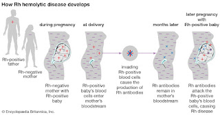

Figure 3.  Illustration of fetal hemolytic disease development

As previously mentioned, individuals with golden blood are able to donate to anyone with common or specific rare Rh system blood types without their blood generating an immune response in the recipient.6 Before Rhnull blood was first discovered it was thought an individual without these antigens would not survive utero.6 Since its discovery, golden blood has been sought after by researchers and physicians alike for its rare properties, however, in 2017 there were only 9 active donors.11 This makes finding a single bag of the blood extremely difficult which has made blood transfusions for these patients complicated and slowed the progression of studies on the blood.11  Despite this, with all of its very special characterizations, these individuals’ blood holds an extraordinary place in the healthcare and scientific communities.6

References:

1. Rh Factor. American Pregnancy Association. https://americanpregnancy.org/pregnancy-complications/rh-factor/. Published October 9, 2019.
2. Journal Of Pakistan Medical Association. JPMA. https://jpma.org.pk/article-details/2399.
3. Stomatocyte. Stomatocyte - an overview | ScienceDirect Topics. https://www.sciencedirect.com/topics/medicine-and-dentistry/stomatocyte.
4. Glossary: Alloantibody - Blood Bank Guy Glossary. Blood Bank Guy. https://www.bbguy.org/education/glossary/gla17/.
5. Blood: the basics. Professional Education. https://professionaleducation.blood.ca/en/transfusion/publications/blood-basics. Published February 11, 2019.
6. Bailey P. Wellcome. The man with the golden blood. https://mosaicscience.com/story/man-golden-blood/.
7. Hemolytic Anemia. Johns Hopkins Medicine. https://www.hopkinsmedicine.org/health/conditions-and-diseases/hemolytic-anemia.
8. Osmotic fragility test: MedlinePlus Medical Encyclopedia. MedlinePlus. https://medlineplus.gov/ency/article/003641.htm.
9. Cartron, Jean-Pierre. "Rh-deficiency syndrome." The Lancet 358 (2001): S57.
10. Hemolytic Anemia. National Heart Lung and Blood Institute. https://www.nhlbi.nih.gov/health-topics/hemolytic-anemia.
11. Rhnull, the Rarest Blood Type on Earth, Has Been Called the "Golden Blood". Curiosity.com. https://curiosity.com/topics/rhnull-the-rarest-blood-type-on-earth-has-been-called-the-golden-blood-curiosity/.

By Cheyenne Cook, A Master’s of Medical Sciences Student at the University of Kentucky

Monday, November 25, 2019

What a Headache!


Migraines are the most common cause of neurological disability in the world affecting nearly 15% of the global population. Some migraines initially present with prodromes or warning signs, which may include: Fatigue, euphoria, depression, irritability, food cravings, constipation, neck stiffness, increased yawning, and/or abnormal sensitivity to light, sound, and smell.2 The prodrome phase is often followed by the aura phase. Auras are transient neurological pathologies that manifest as scintillating light and blurred field of vision and progress to loss of peripheral vision creating tunnel vision. Not all who suffer from migraines get auras, though prodromes are very common. The headache phase, the final phase, proceeds the aura when present, typically pulses; this increases intracranial pressure, and may present in association with nausea, vomiting, and abnormal sensitivity to light, noise, and smell. It may also be accompanied by abnormal skin sensitivity and muscle tenderness.2
Adding another layer to the difficulty of this disease is the fact that it can worsen over time as it transitions from acute to chronic manifestation. Multiple factors that contribute to this chronification are shown in Figure 1. Migraines should be viewed as a complex disorder with a strong genetic basis involving cortical, subcortical and brainstem regions that account for the pain and wide variation of symptoms1. The extent and diversity of symptoms strongly suggest that migraines are more than just headaches. Treatment options are complex, broad, and often countercurrent with rational thinking; successful pharmacological treatment for one person, may be a trigger and detriment for another. The diversity in symptoms of migraines is reflected in the diversity of pharmacological treatments for them; and largely follow one of two paths: Prophylactic or therapeutic. Two of the most promising pharmacological treatments of migraines, Triptans and Onabotulinumtoxin A, will be discussed in more detail below.

Figure 1: Contributing Factors in Migraine Chronification (3)


                The Triptans, introduced nearly 30 years ago, are still considered the gold standard of migraine treatment. These drugs are selective serotonin receptor agonists, and because of their effectiveness have largely replaced older drugs in this category like the ergot derivatives. The Triptans are potent vasoconstrictors and are thought to target 5-HT1B/1D receptors (trigeminovascular afferents and trigeminal nucleus caudalis), to inhibit the release of neurotransmitters.4 The pharmacokinetic properties of the most prominent Triptans are shown in Figure 2. All of the drugs in this class are indicated for the acute treatment of mild to severe migraines that respond poorly to nonsteroidal anti-inflammatory drugs with or without the addition of additional analgesics, and mild to severe migraines in patients with contraindications, intolerance or hypersensitivity to other analgesics. Most Triptans are available in various formulations including: Subcutaneous injection, oral tablet, nasal spray, and oral dispersible tablet. The choice of formulation is determined by a number of patient factors that include headache features, side effect preference, convenience and cost. The single patient’s response to a Triptan cannot be predicted, but most show highest effectivity if they are taken at the very onset of the headache phase.4 All of the Triptans show similar side effects: Paresthesia, flushing, tingling of hands and feet, and mild, fleeting chest pressure. 1 in 1,000,000 patients experience cardiovascular complications of arrhythmia, stroke, and heart attack.

Figure 2: Pharmacokinetics of Triptans (4)

            Central and peripheral nerve sensitization occurs during the progression of migraines. This refers to the nerve response threshold decreasing and their response magnitude increasing. It stands to reason then – by paralyzing the muscle at the neuromuscular junction, one can mitigate some of the effects of migraines. That is precisely the mechanism behind the use of Botulinum neurotoxins (BoNTs). First used as a cosmetic enhancement to prevent wrinkles, BoNTs are now known to be effective in preventing migraines. BoNTs are produced by seven serotypes (A-G) of bacillus bacteria, Clostridium botulinum, and consist of a heavy chain and a light chain. The heavy chain facilitates the uptake of the whole molecule into the cytosol, and the light chain then cleaves soluble N-ethylmaleimide-sensitive factor attachment protein receptor (SNARE) complex in the motor neurons.SNARE molecules are critical to this mechanism because they fuse synaptic vesicles containing neurotransmitters like acetylcholine with the pre-synaptic membrane; which results in the release of the neurotransmitter back into the pre-synaptic cell; without transmission to the post-synaptic cell, paralysis will occur. Onabotulinumtoxin A (onaBoNTA), approved by the FDA in 2010, represents the only agent specifically approved for the prevention of chronic migraines. In addition to modulating the function of motor neurons, uptake of onaBoNTA in sensory neurons that innervate the skin and muscles is thought to inhibit the release of inflammatory mediators at several sites within the sensory neuron.5 OnaBoNTA is administered in accordance with the PREEMPT regimen at 12-week intervals. 31 sites across frontal, temporal, occipital and neck muscles are injected with 5 units per injection, totaling 155 units; the injection scheme can be seen in Figure 3. A Botox unit is a measure of a defined amount of biological activity and corresponds to a fixed number of botulinum toxin molecules. 


Figure 3: Injection Sites for OnaBoNTA (9)


  Migraines are highly heterogeneous, and their treatment should be tailored to the individual patient. The variability in migraine etiology informs the numerous pharmacological treatments; Triptans and OnaBoNTA only scratch the surface of available options. After explaining the diagnosis, the provider should encourage the patient to actively participate in assessing his/her lifestyle and behaviors; diet, sleep, stress and exercise, may all contribute to the individual’s condition and subsequent response to pharmaceutical intervention.

References

1.      Puledda, Francesca et al. “An update on migraine: current understanding and future directions.” Journal of neurology vol. 264,9 (2017): 2031-2039. doi:10.1007/s00415-017-8434-y
2.      Burstein, Rami et al. “Migraine: multiple processes, complex pathophysiology.” The Journal of neuroscience : the official journal of the Society for Neuroscience vol. 35,17 (2015): 6619-29. doi:10.1523/JNEUROSCI.0373-15.2015
3.      May, Arne, and Laura H. Schulte. “Chronic Migraine: Risk Factors, Mechanisms and Treatment.” Nature News, Nature Publishing Group, 8 July 2016, https://www.nature.com/articles/nrneurol.2016.93.
4.       Antonaci, Fabio et al. “Recent advances in migraine therapy.” SpringerPlus vol. 5 637. 17 May. 2016, doi:10.1186/s40064-016-2211-8
5.      Gooriah, Rubesh, and Fayyaz Ahmed. “OnabotulinumtoxinA for chronic migraine: a critical appraisal.” Therapeutics and clinical risk management vol. 11 1003-13. 29 Jun. 2015, doi:10.2147/TCRM.S76964
6.      Silberstein, Stephen D., et al. “OnabotulinumtoxinA for Treatment of Chronic Migraine: PREEMPT 24-Week Pooled Subgroup Analysis of Patients Who Had Acute Headache Medication Overuse at Baseline.” Journal of the Neurological Sciences, vol. 331, no. 1-2, 2013, pp. 48–56., doi:10.1016/j.jns.2013.05.003.
7.      Goadsby, Peter J et al. “Pathophysiology of Migraine: A Disorder of Sensory Processing.” Physiological reviews vol. 97,2 (2017): 553-622. doi:10.1152/physrev.00034.2015
8.      Akerman, Simon, et al. “Current and Novel Insights into the Neurophysiology of Migraine and Its Implications for Therapeutics.” Pharmacology & Therapeutics, vol. 172, 2017, pp. 151–170., doi:10.1016/j.pharmthera.2016.12.005.
9.      “Botox For Migraine.” MigrainePal, 21 Jan. 2019, https://migrainepal.com/botox-for-migraine/.
10.  Sutherland, Heidi G., et al. “Advances in Genetics of Migraine.” The Journal of Headache and Pain, vol. 20, no. 1, 2019, doi:10.1186/s10194-019-1017-9.
By Danny Craig, A Master’s of Medical Science Student at the University of Kentucky

Friday, November 22, 2019

Caloric Restriction: Can it help you increase your rate of living?

          In the past few years, dieting has become such an important part of society. Society is addicted to losing weight, gaining muscle, attaining better health, and trying to live longer. In Caloric restriction (CR),  in the absence of malnutrition, has shown to extend lifespan in many animal models and decrease age-related oxidative stress in multiple tissues & organs 1. Furthermore, recent studies show CR extends the lifespan and reduces a number of adverse effects associated with aging. The principal theory on caloric restriction is a combination of “rate of living” and “oxidative stress” theories, which emphasize the importance that metabolic rate and reactive oxygen species have on lifespan 4. The ‘rate of living’ theory claims that the higher an organism’s metabolism, the shorter its life span, thus explaining the effects of caloric restriction on longevity 4. The ‘oxidative stress’ theory claims that caloric restriction reduces oxidative stress, which in turn reduces damage to tissues and organs (Figure 1) 3,4.  Researchers in the caloric restriction field have examined how age-related changes in physiological and endocrine systems play a role in metabolic functions, which may be correlated with health and lifespan benefits 4.


Figure 1.  CR enhances resting energy efficiency to decease systemic oxidative damage (3).

An example of ongoing work investing how CR may benefit overall health is a study by Redman et al 2. Here, it was hypothesized that maintaining patients on CR diets for a period of two years would reduce energy expenditure (EE) of endocrine mediators and oxidative stress in energetically active tissues 2. Finally, the authors conducted this study to provide scientific validity for the biological aging hypothesis; which explains that a prolonged use of CR diet enhances energy efficiency, reduces reactive oxygen species, and reduces oxidative damage to tissues and organs 3.

The researchers recruited 53 people, of which only 19 people were assigned to the control group and 34 people were randomized to the caloric restriction group 3. In order to see how CR plays a role the amount of weight each group lost was examined 3. The CR group lost about 9.4 kg on average and a 16.5% reduction in energy intake 3In year two the CR group only maintained their weight loss and had a 14.8% reduction in energy intake (Figure 2) 3. The weight loss in the CR group was primarily fat mass loss, but some fat-free mass loss was observed as well 3This study explains the idea that changes in metabolic adaptation of energy expenditure show reductions in oxidative stress 3. This implies that metabolic adaptation may be related to energy conservation, and in turn may delay biological aging, which in turn enhance lifespan 3.
 Fat mass loss was observed in the CR group and to understand how this effects energy metabolism, aging, and metabolic changes 3. To examine these effects the researchers attained blood samples from both groups 3. The blood samples were tested for T3, T4, TSH, leptin, and insulin 3.  It was found that leptin levels drastically decreased in the CR group and the control group had no changes 3. When examining T3 and T4 levels the CR group had significant reductions in the concentration of these hormones over the two years 3. When examining TSH there was not a considerable impact on the thyroid axis and does not explain how TSH plays a role in mediating metabolic adaption 3.  Finally, insulin concentration were significantly lowered in the CR group at year one but not in year two, and in the control group insulin levels were maintained for both years 3. 


Figure 2.  Calorie Restriction and Change in Body Composition.  Percent of calorie restriction (A) achieved after 1 and 2 years of calorie restriction and the resulting change in fat mas (FM) and fat-free mass (FFM) (B).  N=53: 34 CR, 19 controls.  The p value for statistically significant treatment group effects, adjusted for multiple comparisons, is shown.  The changes in weight, FM and FFM were all significantly different between the CR and control group (p<0.0001 for all, treatment main effect)(3).
Oxidative stress was measured to understand how the biological aging hypothesis. The participants gave urine samples, which were used to measure urinary 2,3-dinor-iPF(2α)-II by liquid chromatography to see oxidative stress 3The urinary F2-isoprostane excretion showed a significant reduction in the CR group but no changes in the control group 3When analyzing the urinary data, it was found that urinary 2,3-dinor-iPF(2α)-II concentrations where significantly lower for the CR group than the control group at year two 3These changes in the urinary 2,3-dinor-iPF(2α)-II concentration show a drop in oxidative stress, but these findings need to be studied long-term 3.
Redman et al, explain that patients who were put on a caloric restriction diet for two years had an impact on biological aging 3. This study shows that the use of CR does have some beneficial aspects to human life span due to efficiency in energy metabolism, fat loss, and reducing systemic oxidative stress. CR’s effectiveness is important as it indicates weight loss to be safe and tolerable, as well as a reduction in systemic oxidative stress to live longer. Overall, caloric restriction is important for people’s health, people’s lifespan benefits, and allow people to live longer.  

By Cocanut Suhail, a Master's of Medical Science Student at the  University of Kentucky

References:

1.      Il’yasova, D., Fontana, L., Bhapkar, M., Pieper, C., Spasojevic, I., Redman, L., Das, S.K., Huffman, K., and Kraus, W. (2018). Effects of 2 years of caloric restriction on oxidative status assessed by urinary F2-isoprostanes: The CALERIE 2 randomized clinical trial. Aging Cell. 17: e127109.

2.      Ravussin, E., Redman, L., Rochon,J., Das, S.K., Fontana, L., Kraus, W.,  Romashkan, S., Williamson, D., Meydani, S., Villareal, D.T., Smith, S., Stein, R., Scott, T., Stewart, T.,Saltzman, E., Klein, S., Bhapkar, M., Martin, C., Gilhooly,  Holloszy, J., Hadley, E., Roberts, S., and Kritchevsky, S.  (2015). A 2-Year Randomized Controlled Trial of Human Caloric Restriction: Feasibility and Effects on Predictors of Health Span and Longevity, The Journals of Gerontology: Series A. 70 (9), 1097–1104.

3.      Redman, L. M., Smith, S. R., Burton, J. H., Martin, C. K., Il'yasova, D., & Ravussin, E. (2018). Metabolic slowing and reduced oxidative damage with sustained caloric restriction support the rate of living and oxidative damage theories of aging. Cell Metabolism. 27(4), 805-815.


4.      Speakman, J., and Mitchell, S. (2011). Caloric restriction. Molecular Aspects of Medicine. 32: 159-221.


Thursday, November 21, 2019

To Die or not to Die


        Everything and everyone that is living has a limit to life, down to the single unit of living things our cells go through the same fate, death. During development, many regulatory mechanisms help direct and promote either cell differentiation and growth or the opposite cell death. Imagine trillions and trillions of cells accumulating.  If they  happened to live forever, it would cause many mechanisms that regulate physiological homeostasis to fail. We would all have high blood pressure and massive edema and eventually death if certain cells did not have a regulatory control mechanism. Cell death is necessary and specifically controlled. Three  main ways a cell can undergo cell death are apoptosis, necrosis, or autophagy. All three of these play vital roles in controlling many processes in development, the development and progression of major diseases and basic, daily functions. These cell death processes are also essential therapeutic targets to treat disease a variety of disease states.  Promoting death of cancer cells or preventing death of neurons  in neurodegenerative diseases like  Parkinson’s disease or  other tauopathies may be beneficial.
        During the development of cancer, defects in apoptosis pathway results in an accumulation of cells leads formation of a tumor. A crucial step in apoptosis involves activation of caspases (i.e., caspases 3, 7 and 9) by signaling and activating MEK through dimerization of the receptor which activates the ERK and MAPK which can phosphorylate caspase 9. Another way to phosphorylate caspase 9 is through mitosis and buildup of CDK1 and Cyclin B. Apaf-1 is also needed to form a complex with caspase 9 in order to activate caspase 3 and 7. This occurs by the BH3 pro cytochrome c activator and Bcl-2 a anti cytochrome c activator to bind to Apaf-1. After Caspases 3 and 7 are activated, apoptosis can be initiated (Fig. 1).  Overall apoptosis does not cause toxic build up at the site of cell death and has a very rapid turnover.

Figure 1: Caspase-9 is activates by formation of the apoptosome and by forming a multimeric complex with Apaf-1. When cytochrome c is released. This can be enhanced by proapoptotic signal BH3 or inhibited Bcl-2 an antiapoptotic protein class2
        Like apoptosis, necrosis, is also a clinically important process of cell death.  Necrosis is involved in tissue damage that occurs during traumatic brain injury, myocardial infarctions, strokes and some liver diseases.  While it is also initiated following DNA fragmentation and dysregulate mitochondrial function, it fails to be subject to processed by lysosomal degradation.  Instead, necrotic cells spill their  toxic contents out into the extracellular space aiding in an increase in tissue damage and inflammation brought on by cytokines. Assays to distinguish different forms of cell death are used, including use of  a deoxynucleotidyl transferase-mediated biotin-dUTP nick-end labeling assay to detect DNA fragmentation3. Ischemia and hypoxia are important triggers of cell necrosis. While necrosis also has tightly regulated features, targeting these has limited  therapeutic value, since the damage is already done such as many agents in Spinal cord injuries have been related to inhibiting further necrosis and reducing inflammation through the process of apoptosis. Energy starvation or oxygen starvation, ROS production can generate pro-necrotic signals which consist of inflammatory mediators and their ligands. Cellular calcium overload accompanies depletion of intracellular adenosine triphosphate (ATP), since calcium pumps that sequester cytosolic calcium are energy dependent. Calcium overload is a potent inducer of cell death3. An indirect therapy suggests that activating protein kinase (MAP) turns necrosis signaling into stress activates induced apoptosis. Inhibition of p38 is another way to also reduce cellular necrosis. A common pathology associated with necrosis is pyknotic cells which degrade and increase toxicities to surrounding cells (Fig 1). This displays the importance of a balancing act of pro and antiapoptotic signals.
Figure 2: Distinctive features of the CSC (cancer stem cell) niche, including hypoxia, reduced nutrient availability and acidity (H+), promote high levels of basal autophagy in CSCs4. This figure explains the Basal autophagy pathway in different cancer cells that are self-renewing rapidly. These cells can undergo basil autophagy if high acidity, low glucose or oxygen is present acutely, this initiates different signaling mechanisms such as inhibition of ROS build up, migration leading to metastasis. And even further cell differentiation.
        Autophagy is the third natural cell death process and occurs when the body undergoes extreme change in pH, glucose deficiency, and hypoxic conditions. This pathway has the most potential for being targeted for the development of new therapies to treat  cancer. Autophagy can occur during fasting and inhibit production of reactive oxygen species and cell death.  Mechanistically it is controlled/initiated by the mTOR pathway and activates through the ATG/ULK1 and Class III PI3K complex. Next the autophagosome formation is meditated by ATG7,10,5,12. It them fuses with a lysosome degrades the waste into amino acids, lipids and nucleotides to be recycled to a new cell system. In a way autophagy cleanses the body to a degree and is a reset for cellular processes and resulting in a less toxic and efficient/stable cellular enviorment5.
        Taking all cell death pathways into account, we can better understand the pros and cons of cell death mechanisms therapeutically  for each specified disease whether its cancer and for example Hotchkiss et al classified that decreased apoptosis is associated with diseases in over half of neoplasms. Enhancing apoptosis TP53 guardian can reduce the risk of cancer by 8-fold. Also stated in a table Leukemia and multiple myeloma and colorectal cancers have been treated with obatoclax which induces apoptosis by inhibiting antiapoptotic Bcl-2 family members. Or activates death receptors for treating Hodgkin’s lymphoma6. Another example is giving hydroxychloroquine to inhibit autophagy in breast cancer and prostate cancer, which can ultimately inhibit the progression and metastasis of early stages of these cancers. It is important to look at death as good at times and appreciate the amount of interplay of genes and signaling cascades there are for normal biochemical processes within our complex systems. This all concludes the importance apoptosis is for normal function and health to our biological systems.

References:
(1)    Reed, J. C. (2000). Mechanisms of Apoptosis. The American Journal of Pathology, 157(5), 1415–1430. doi: 10.1016/s0002-9440(10)64779-7
(2)    Allan, Lindsey & Clarke, Paul. (2008). Allan LA, Clarke PRA mechanism coupling cell division and the control of apoptosis. SEB Exp Biol Ser 59: 257-265. SEB experimental biology series. 59. 257-65.
(3)    Szabó, C. (2005). Mechanisms of cell necrosis. Critical Care Medicine, 33(Suppl). doi: 10.1097/01.ccm.0000187002.88999.cf
(4)    Boya, P., Codogno, P., & Rodriguez-Muela, N. (2018). Autophagy in stem cells: repair, remodelling and metabolic reprogramming. Development, 145(4). doi: 10.1242/dev.146506
(5)    Yang, Z., & Klionsky, D. J. (2009). An Overview of the Molecular Mechanism of Autophagy. Current Topics in Microbiology and Immunology Autophagy in Infection and Immunity, 1–32. doi: 10.1007/978-3-642-00302-8_1
(6)    Hotchkiss, R. S., Strasser, A., McDunn, J. E., & Swanson, P. E. (2009). Cell death. The New England journal of medicine361(16), 1570–1583. doi:10.1056/NEJMra0901217

Amal Agarwal University of Kentucky MSMS student Class of 2019


Friday, November 8, 2019

Biologic Drugs: From Crohn’s Disease to Cancer


            Biologics are a wide class of drugs that in recent decades, have become incredibly common and desirable due to their specificity and subsequent decreased potential for side effects. All biologics originate from or are modified forms of biomolecules. In contrast to traditional small molecule drugs such as lipitor or aspirin, biologics are quite large and are substantially more specific in the treatment of small groups of diseases through targeted modification of cellular pathways (Chan and Chan, 2017). This is by design, as most new biologic drugs are synthetic and rely on exploiting the principles of established physiological actions. Many believe that the healthcare industry is only just beginning to understand and utilize this powerful class of drugs and in fact they may allow for the treatment of many diseases that current lack effective therapies (Riley, 2018).
            One common example of a disease that is preferentially treated with biologic drugs is Crohn’s disease or CD for short. CD is a chronic inflammatory disease of the gastrointestinal tract that is caused by dysregulation of inflammatory pathways within the walls of the digestive system (Binion, 2019). This disease has been on the rise in recent decades.  It is now estimated that more than 1.6 million individuals have been diagnosed with CD in the United States (Shivashankar et al, 2017). Prior to the advent of biologics, CD was treated in much the same way as other inflammatory conditions, with repeated administration of corticosteroids and eventual surgery. The corticosteroids would down regulate and maintain low levels of inflammation, while surgery would remove the areas of the GI tract that were inevitably destroyed by the disease (Binion, 2010). It is not difficult to imagine that the prospects for life with CD were not great at that time. Now, CD is treated using biologics such as infliximab and adalimumab which modulate tumor necrosis factor or TNF which is a major cytokine that is crucial for maintenance of the inflammatory pathway. By binding and removing TNF, inflammation is halted in patients with CD and remission is commonly achieved and maintained (Jauregui-Amezaga et al, 2017).
            Despite the success of biologics, however, it is not uncommon for some patients to become unresponsive to the drug or to develop resistance over time (Riley, 2018). Because of this, a patient may have to switch between drugs throughout the course of their lifetime. Interestingly, more and more biologics are made each year, and the newest generation of biologics are designed to be more efficient and less likely to lose effectiveness. One such CD drug is ustekinumab which was originally used for psoriatic arthritis but is now evaluated for other applications like CD. In CD, ustekinumab, a monoclonal antibody, targets the p40 subunits of cytokines IL-12 and IL-23 to downregulate the immune response and shut down inflammation. Of note, these pathways also prohibit TNF and their downstream activity in the Th17 and Th1 response pathways (Jauregui-Amezaga et al, 2017).

Figure 1. An illustration of the action of ustekinumab on p40 and cytokine activation (Jauregui-Amezaga et al, 2017)

            Many patients with CD benefit from biologics, but the applications of these drugs are far more diverse with uses in treating several forms of arthritis, asthma, lupus, diabetes, heart attack, cystic fibrosis, osteoporosis, and several more. Several more diseases have the potential to benefit from future biologics that are not even yet made such as multiple sclerosis and cancer (Riley, 2018). Cancers are of particular interest because of the horrible side effects that are typically associated with chemotherapy, the current standard of cancer treatment. The principles that are guiding research efforts into the development of biologic cancer drugs rely on targeting markers specific to a cancerous cell using antibodies.  These antibodies may then arm immune cells to destroy cancer cells directly using the host immune system. They can also act to destroy cancer cells by introducing cytotoxic agents by binding a target marker and entering the cell with the toxin (NIH National Cancer Institute, 2018). Regardless, this could be far more specific than traditional chemotherapy that indiscriminately destroys cells and tissues leading to side effects like hair loss, soft tissue damage, and neuropathies.

Figure 2. An illustration of endocytosis of biologic drugs conjugated to cytotoxic agents in cancer cells (Riley, 2018).
             There is now a sort of mad dash to develop these drugs because of how potentially lucrative producing biologics can be. More than 37% of net drug spending since 2014 was on biologics, and more than 93% of the drug market growth can be attributed to an increase in their sales and production (Forbes, 2019). It is quite easy to forecast that the market will continue to demand more biologics as time goes on. More importantly, many diseases that could not be managed before will likely have biologics developed for them to improve the quality of life for patients and enrich our knowledge about disease processes and treatment at large. As such, the healthcare industry and patients will no doubt continue to benefit from biologic drugs as they evolve and improve.

By Bradley Wright, Master's of Medical Science Student, University of Kentucky

References:

Binion, D. G. (2010). Biologic Therapies for Crohn's Disease Update from the 2009 ACG Meeting. Gastroenterology and Hepatology6(1), 4–16. Retrieved from https://www.ncbi.nlm.nih.gov/pmc/articles/PMC2886448/

Riley, M. (2018, March 13). The ever-increasing attraction of biologics. Retrieved October 26, 2019, from https://www.chemistryworld.com/health-tech/the-ever-increasing-attraction-of-biologics/3008719.article.
Chan, J. C., & Chan, A. T. (2017). Biologics and biosimilars: what, why and how? ESMO Open2(1). doi: 10.1136/esmoopen-2017-000180
Jauregui-Amezaga, A., Somers, M., Schepper, H. D., & Macken, E. (2017). Next generation of biologics for the treatment of Crohn’s disease: an evidence-based review on ustekinumab. Clinical and Experimental GastroenterologyVolume 10, 293–301. doi: 10.2147/ceg.s110546
NIH National Cancer Institute. (2018, April 26). Biological Therapies for Cancer. Retrieved October 26, 2019, from https://www.cancer.gov/about-cancer/treatment/types/immunotherapy/bio-therapies-fact-sheet.
Forbes. (2019, July 3). Biologic Medicines: The Biggest Driver Of Rising Drug Prices. Retrieved October 26, 2019, from https://www.forbes.com/sites/theapothecary/2019/03/08/biologic-medicines-the-biggest-driver-of-rising-drug-prices/#61afc63718b0.
Shivashankar, R., Tremaine, W. J., Harmsen, W. S., & Loftus, E. V. (2017). Incidence and Prevalence of Crohn’s Disease and Ulcerative Colitis in Olmsted County, Minnesota From 1970 Through 2010. Clinical Gastroenterology and Hepatology15(6), 857–863. doi: 10.1016/j.cgh.2016.10.039


A Way Out


A majority of our life is defined by our choices. For the most part, we have the power to decide everything from what we want to eat today to what we want to spend the rest of our lives doing. But what about those life-altering decisions that have already been made for us, sometimes even before we are born? Is it just the luck of the draw? Should we just accept the cards we were dealt with or is there something we can do to  change the course of our lives?

        For years, many have succumbed to incurable diseases because the therapies used to treat the disease were anything but sufficient. People worldwide suffer from inherited disorders that can cost them later in life and even impact the future of generations to come. One such disease is hypertrophic cardiomyopathy (HCM). HCM is a genetic disorder that displays an autosomal dominant inheritance pattern1. According to the National Human Genome Research Institute, autosomal dominant explains how certain traits are inherited. Autosomal signifies that the trait acquired is from a non-sex chromosome; thus, for humans, autosomal diseases are derived from chromosomes 1 through 22. Furthermore, dominant explains how many copies of the mutated gene are required for the disease phenotype. If it is dominant disorder, then only one copy of the mutated gene will display the disease phenotype. Other examples of autosomal dominant diseases are Huntington disease and Marfan Syndrome2.

        Therefore, in cases of familial hypertrophic cardiomyopathy, it only takes one mutated
gene to dictate what the rest of your life will look like. HCM is depicted by the thickening of the left ventricle of the heart, lowered chamber capacity, and an enlarged heart1. The muscle walls of heart thicken which restricts blood flow causing the ventricle to work harder to pump blood to the rest of the body4. The prevalence of HCM is estimated to occur in around 1 out of 625 people to 1 in 344 people1. The European Society of Cardiology suggests that a heart wall thickness greater than or equal to 15 mm is indicative of HCM3.  HCM is a hereditary disorder which affects approximately 0.2% of the population worldwide1. Common symptoms of HCM include dizziness, fainting, fatigue, chest pain, shortness of breath, and arrhythmias which can lead to death7.
        While a number of different mutational events can occur, the most prevalent, accounting for up to 40% of HCM cases involve mutations in a gene called MYBPC3.  This gene is responsible for creating the cardiac myosin binding protein C, a cardiac protein, and is typically inherited from at least one parent8. Treatment of patients who have MYBPC3 mutations involve regular monitoring and medications and to open heart surgery or an implantable cardioverter-defibrillator (ICD)5. While these treatments options will only alleviate symptoms and are temporary solutions, there is no cure presently. The survival rate of people with HCM is 98% after 1 year, 94.3% after 3, and 82.2% after 56. While HCM is not necessarily a death sentence, a person’s mortality is always looming over their shoulder. People should not have to live in fear, wondering which day will be their last. Perhaps with genome editing, they won’t have to anymore.

        In a 2017 study conducted by Ma et al, the effects of genome editing to correct germline mutations that cause HCM were explored. Here, healthy gamete donors who had homozygous and heterozygous heritable MYBPC3 mutations were used. They used CRISPR-Cas9 to specifically target the heterozygous MYBPC3 mutation in embryos prior to implantation8. CRISPR-Cas9 is a genome editing technology that allows for an organism’s DNA to be altered. The technology involves creation of a small piece of RNA which contains a guide sequence that attaches to a specific portion of the DNA. That RNA piece will also bind to an enzyme called Cas9. Essentially, the RNA recognizes the portion of DNA which needs to be removed from the genome and Cas9 will cut that DNA at the specific location. After the DNA is cut, a customized DNA sequence will replace the sequence that was removed from the genome9. Therefore, this technology is especially promising for individuals with mutations that cause incurable diseases. 

        Ma et al. explains that homologous direct repair (HDR) is necessary for gene correction. They found that the double strand breaks in the human gametes and zygotes were fixed by using an internal HDR mechanism by using a wild-type allele as a template. However, in induced pluripotent stem cells, the rate of HDR was much lower indicating that DNA damage response system behaved differently in gametes and embryos. One problem, though, is that , there are still off target effects like non-homologous end joining induced indels that need to be controlled for and understood8. We are not close to where we need to be, but this is one step to finding the answer. Undoubtedly, there are many challenges to using gene therapy. The first of which is limited sample size and the difficulty in setting clear boundaries10. Furthermore, editing the germline can cause intended consequences to future generations. Although there is heavy criticism and controversy surrounding this study, I am hopeful for the future. Right now, CRISPR-Cas9 has only been tested on animal models and embryos, but in the future, I expect to see this technology being used in humans who discover their diseases later in life.  After more testing, the goal is to help those who are victims to genetic mutations and allow them to live disease free. If this expands to humans and is successful, individuals will no longer have to succumb to the whimsy of their genes.

By Rachel Crasta, Master of Medical Sciences Student, University of Kentucky

References:

1.      Marian, Ali J., and Eugene Braunwald. “Hypertrophic Cardiomyopathy.” Circulation Research, vol. 121, no. 7, 2017, pp. 749–770., doi:10.1161/circresaha.117.311059.
2.      “Autosomal Dominant.” Genome.gov, NIH, www.genome.gov/genetics-glossary/Autosomal-Dominant.
3.      Authors/Task Force Members, Elliott PM, Anastasakis A, Borger MA, Borggrefe M, Cecchi F, Charron P, Hagege AA, Lafont A, Limongelli G, Mahrholdt H, McKenna WJ, Mogensen J, Nihoyannopoulos P, Nistri S, Pieper PG, Pieske B, Rapezzi C, Rutten FH, Tillmanns C, Watkins H. 2014 ESC guidelines on diagnosis and management of hypertrophic cardiomyopathy: the task force for the diagnosis and management of hypertrophic cardiomyopathy of the european society of cardiology (ESC). Eur Heart J. 2014;35:2733–2779. doi: 10.1093/eurheartj/ehu284.
4.      “Hypertrophic Cardiomyopathy.” Www.heart.org, www.heart.org/en/health-topics/cardiomyopathy/what-is-cardiomyopathy-in-adults/hypertrophic-cardiomyopathy.
5.      Mcnamara, James W., et al. “MYBPC3 Mutations Are Associated with a Reduced Super-Relaxed State in Patients with Hypertrophic Cardiomyopathy.” Plos One, vol. 12, no. 6, 2017, doi:10.1371/journal.pone.0180064.
            6.    Liu, Qun, et al. “Survival and Prognostic Factors in Hypertrophic Cardiomyopathy: a Meta-  
                  Analysis.” Nature News, Nature Publishing Group, 20 Sept. 2017,                          
                  https://www.nature.com/articles/s41598-017-12289-4.
7.      “Hypertrophic Cardiomyopathy.” HIE Multimedia - Hypertrophic Cardiomyopathy, slu.adam.com/content.aspx?productId=117&isArticleLink=false&pid=1&gid=000192.
8.      Ma, Hong, et al. “Correction of a Pathogenic Gene Mutation in Human Embryos.” Nature, vol. 548, no. 7668, 2017, pp. 413–419.,doi:10.1038/nature23305.
9.      “What Are Genome Editing and CRISPR-Cas9? - Genetics Home Reference - NIH.” U.S. National Library of Medicine, National Institutes of Health, ghr.nlm.nih.gov/primer/genomicresearch/genomeediting.
10.  Papasavva, Panayiota, et al. “Rare Opportunities: CRISPR/Cas-Based Therapy Development for Rare Genetic Diseases.” Molecular Diagnosis & Therapy, vol. 23, no. 2, 2019, pp. 201–222., doi:10.1007/s40291-019-00392-3.


Monday, November 4, 2019

In Mice and Men (and Women): Prostate Drug, Terazosin to Slow Progression of Parkinson’s Disease


        In 1817, English physician James Parkinson published “An Essay on the Shaking Palsy,” which was the earliest known comprehensive explanation of Parkinson’s Disease (PD). Over 200 years later the underlying basis causing PD remains a mystery. However, research suggests that PD is a consequence of the complex, multifaceted relationship between several environmental and genetic components.1 Leucine-rich repeat kinase 2 (LRRK2) is a kinase enzyme encoded by the LRRK2 gene. Mutations or variations of the LRRK2 gene have been deemed as the principle factor manipulating the genome in most cases of familial PD, and some cases of sporadic PD.1 Hallmark signs and symptoms of PD (Figure 1.) are typically motor-related, for instance, the most common PD motor symptoms include bradykinesia, rigidity, resting tremor, stooped posture/unstable balance.1,2
Figure 1. Hallmark Symptoms and Signs of PD.2

        The basal ganglia system plays a major role in the initiation of movements (motor planning) and muscle tone. Two of the major nuclei found in the basal ganglia are the substantia nigra and the striatum. The striatum is further divided into three subsections, the caudate nucleus, putamen, and nucleus accumbens. Together, the caudate nucleus and putamen form the dorsal striatum. The caudate nucleus receives afferent signals from the prefrontal cortex, and it is responsible for cognitive function and regulating eye movements. On the other hand, the putamen receives afferent inputs from the motor cortex and the substantia nigra and it controls motor function, specifically voluntary movement. Another key nucleus of the basal ganglia system is the substantia nigra. Neurons of the substantia nigra use dopamine as a neurotransmitter, giving this area its unique distinguishing appearance.
        The substantia nigra appears unusually dark even in the absence of stain; this distinctive characteristic is a result of neuromelanin. Neuromelanin is a dark pigment formed during dopamine metabolism and is continuously produced by the densely packed dopamine neurons of the substantia nigra. These dopaminergic neurons are projected from the substantia nigra to the putamen region of the striatum, these afferent fibers are known as nigrostriatal fibers.
        Parkinson’s Disease progressively affects the nervous system, slowly degenerating and destroying dopaminergic neurons in the substantia nigra. As PD advances fewer neurons can be found in the substantia nigra which means that less dopamine is being released into the striatum. The neurodegenerative effects and dysfunction of the nigrostriatal striatal system observed in PD patients are direct consequences of the striatal dopamine-deficiency. 4,5 The absence of striatal dopamine results in the inability to conduct controlled, fine muscle movements.     
         Since there is no definitive diagnostic or imaging test to diagnose PD, diagnosis is made clinically. A clinical diagnosis of PD is only possible once the patient starts to exhibit symptoms of the disease. Once symptoms begin to manifest this indicates that the disease has progressed to a point where over 60% of nigral nerve cells have disappeared. 1 Every case of PD progresses differently, the disease may differ in severity, rate of progression, and an individual may experience different PD symptoms, presentation of neuropathology, age of onset, etc. An example of the average or general progression of PD and the clinical symptoms associated with each stage is shown in Figure 2.
Figure 2. Clinical symptoms associated with PD and general disease progression.3
In many cases, after the patient succumbs to the disease their brain can be examined for conclusive evidence of PD. For example, by immunohistochemically staining the substantia nigra of a PD patient, researchers can establish the extent of neuronal degeneration by comparing the section to the immunohistochemically stained substantia nigra from a healthy patient. 4,5
Unlike the healthy substantia nigra which consists of distinct deeply stained regions, a PD patient’s substantia nigra will show much lighter regions, ultimately allowing researchers to define the disease due to the observed depigmentation (Figure 3).3 
Figure 3. Main neuropathologies observed in immunohistochemical stains of PD patients.3 
In addition to the above-mentioned depigmentation neuropathological hallmark of Parkinson’s Disease is the presence of Lewy Bodies. Lewy Bodies are intracytoplasmic inclusions containing aggregations of the α‑synuclein protein.1,3 Lewy Bodies are known to cause mitochondrial dysfunction by disrupting the electron transport chain which leads to the degradation of dopaminergic neurons in adulthood. Damage to the mitochondria causes diminished ATP levels in PD patients because of their impaired cerebral glucose metabolism and mitochondrial biogenesis. Cellular dysfunctions, including diminished ATP levels and impaired bioenergetics, have been regularly observed in PD patients and may determine the severity and course of the disease.6 As PD progresses, the abnormal deposition and aggregation of the α‑synuclein protein within cytoplasm gradually increase and begin to involve more brain regions.7 Shown in Figure 4 are examples of immunohistochemical staining of three unique Lewy bodies all differing in their morphological characteristics.
Figure 4. Immunohistochemical staining of α -synuclein shows characteristic Lewy bodies.3

    The gold standard for Parkinson’s Disease drug therapy is L-DOPA, also called levodopa. Systemic administration of L-DOPA in PD patients acts to pharmacologically substitute the diminished levels of dopamine in the striatum.1 L-DOPA is the precursor to dopamine in the dopamine synthesis pathway, and unlike dopamine, can cross the blood-brain barrier.1,8,9 While L-DOPA has been critical in reducing the motor dysfunction experienced by patients with PD, this revolutionary breakthrough treatment has been around for over 50 years without being improved or replaced. 8,9
         L-DOPA treatment solely targets the negative consequences of PD on motor function, but it fails to prevent or slow down the degeneration of dopaminergic neurons. Even more alarming is that all current therapies for PD (including L-DOPA) are only focused on regulating and relieving the symptoms, while the underlying, causative neuronal degeneration remain ignored and thus progressive deterioration of the patients’ health still occurs.1 However, a new pharmacologic breakthrough in the treatment of Parkinson’s may be on the horizon.
  This potentially revolutionary therapy for Parkinson’s treatment is not a novel drug, but instead is a “repurposed drug” called of terazosin. Terazosin is normally utilized for treating benign prostatic hyperplasia which is the enlargement of the prostate.10 Terazosin binds to and activates PGK-1 (phosphoglycerate kinase-1), which is the first ATP-producing enzyme in the glycolysis cycle.11 The PGK-1 enzyme is one of only two enzymes in the glycolysis pathway that are capable of generating ATP.11 Terazosin’s interaction with PGK-1 acts to intensify and strengthen the enzyme’s activity resulting in an upsurge of ATP.11
  The application of terazosin for treating PD has been shown using a model of PD that is induced by MPTP. MPTP is a toxin that induces PD symptoms by destroying dopaminergic neurons, inhibiting the ETC in mitochondria, and lowering tyrosine hydroxylase levels (rate-limiting step in dopamine synthesis). Terazosin has proven effective in decreasing neurodegeneration, slightly reestablishing TH and dopamine, as well as, improving motor function all caused by MPTP.
Figure 5. Immunostaining of Striatum. 
Figure 5 illustrates the deep pigmentation characteristic to the striatum which under normal conditions is densely concentrated with neurons. The middle panels representing the striatum when exposed to MPTP in the absence of Terazosin shows a dramatic loss in pigmentation and therefore a loss in neuronal concentration within the striatum. The final two panels show the striatum exposed to MPTP and Terazosin. Due to the presence of Terazosin when MPTP is in the striatum, the neuronal concentration did not significantly deteriorate.10
            Terazosin appears to be an extremely effective treatment for PD; it has the potential to revolutionize the treatment of neurodegenerative diseases. Administering Terazosin, even after the onset of neurodegeneration, has been shown to successfully slow the rate of cell death, as well as, increase tyrosine hydroxylase levels, dopamine content, and motor performance.10
A major benefit of using a drug that is already well established and used clinically to treat other diseases is that the researchers were able to assess the efficacy of Terazosin in humans more easily and effectively. Additionally, they could easily distinguish and test for a Terazosin-induced effect due to the availability of a vast number of human clinical databases.10 Since Terazosin is not a ‘novel’ drug and has been regularly prescribed to many patients, its safety profile is well-documented. However, the researchers’ database analysis was limited to men since Terazosin is typically only prescribed for treatment of benign prostatic hyperplasia, a disease only affecting men. Another potential limitation of Terazosin is the fact that the exact mechanism that Terazosin employs to effect neurons remains unspecified. A final drawback of Terazosin is its tendency to reduce blood pressure, which is a cause for concern in PD patients who may already have low blood pressure.12
The exciting results obtained from the Terazosin study prove that Terazosin has the capability to effectively increase pyruvate levels (a glycolysis product) in the substantia nigra, striatum, and cerebral cortex. The resulting elevation in ATP levels (cellular energy) can alleviate cellular strain to meet energy demands and improve all aspects of neuronal function. Impaired energy metabolism is a common characteristic of PD as well as other neurodegenerative conditions like Alzheimer’s Disease. Therefore, the cellular benefits of Terazosin demonstrate that this drug can be used as a potential treatment for countless other neurodegenerative diseases. These promising results predict Terazosin to be a dominant candidate for clinical trials in PD, with the hope of repurposing this prostate drug to treat this widely prevalent and detrimental disease.

By Niamh Costello, Master's of Medical Science Student at the University of Kentucky 

 References

1.      Grondin, R. (2018). ANA780: Neurobiology of Brain and Spinal Cord Disorders. ANA780: Neurobiology of Brain and Spinal Cord Disorders. Lexington.
2.      Oppenheimer, M. (2018, February 9). New Developments in Parkinson's Disease - TPG, Inc. http://www.tpgonlinedaily.com/new-developments-parkinsons-disease/.
3.      Poewe, W., Seppi, K., & Tanner, C. (2017). Parkinson Disease. Nature Reviews Disease Primers3(17013). doi: doi:10.1038/nrdp.2017.13
4.      Dickson, D., Braak, H., Duda, J., Duyckaerts, C., Gasser, T., Halliday, G., . . . Litvan, I. (2009). Neuropathological assessment of Parkinson's disease: Refining the diagnostic criteria. The Lancet Neurology, 8(12), 1150-1157.
5.      Halliday, G., Holton, M., Revesz, J., & Dickson, L. (2011). Neuropathology underlying clinical variability in patients with synucleinopathies. Acta Neuropathologica, 122(2), 187-204.
6.      Ekstrand, Mats I., Terzioglu, Mügen, Galter, Dagmar, Zhu, Shunwei, Hofstetter, Christoph, Lindqvist, Eva, . . . Larsson, Nils-Göran. (2007). Progressive parkinsonism in mice with respiratory-chain-deficient dopamine neurons. Proceedings of the National Academy of Sciences of the United States of America, 104(4), 1325-1330.
7.      Braak, Heiko, Tredici, Kelly Del, Rüb, Udo, De Vos, Rob A.I, Jansen Steur, Ernst N.H, & Braak, Eva. (2003). Staging of brain pathology related to sporadic Parkinson’s disease. Neurobiology of Aging,24(2), 197-211.
8.      PD Med Collaborative Group. (2014). Long-term effectiveness of dopamine agonists and monoamine oxidase B inhibitors compared with levodopa as initial treatment for Parkinson's disease (PD MED): A large, open-label, pragmatic randomized trial. The Lancet, 384(9949), 1196-1205.
9.      LeWitt, P. A., & Fahn, S. (2016). Levodopa therapy for Parkinson disease: A look backward and forward. Neurology, 86(14_Supplement_1 Suppl 1), S3-S12.
10.  Cai, R., Welsh, M., Liu, L., et al. (2019). Enhancing Glycolysis Attenuates Parkinson’s Disease Progression in Models and Clinical Databases. The Journal of Clinical Investigation. 129(10):4539-4549. https://doi.org/10.1172/JCI129987
11.  Xinping Chen, Chunyue Zhao, Xiaolong Li, Tao Wang, Yizhou Li, Cheng Cao, . . . Lei Liu. (2014). Terazosin activates Pgk1 and Hsp90 to promote stress resistance. Nature Chemical Biology, 11(1), 19-25.
12.  Shugart, J. (2019, September 20). In Mice and Men, Prostate Drug Reportedly Treats Parkinson's Disease. https://www.alzforum.org/news/research-news/mice-and-men-prostate-drug-reportedly-treats-parkinsons-disease